Stages of Eye Examination
How often should I have a general eye exam?
Even in the absence of vision problems, a thorough eye examination every year will allow you to continue to see well and have healthy eyes for many years. Thanks to a comprehensive eye examination, early diagnosis of diseases such as diabetes, brain tumor, AIDS, Alzheimer's disease, hypertension, blood and heart diseases, metabolic disorders, and Parkinson's disease is possible.
-
Visual acuity test
At the first stage of a detailed eye examination, including a visual acuity test, a phoropter is used to check whether the letters are clearly visible or, for people who cannot read, small figures.
This test uses progressively smaller letters and numbers to help identify problems such as farsightedness and nearsightedness.
-
Examination for the need for glasses (refraction test)
The presence of refractive pathologies such as hypermetropia, myopia, and astigmatism is checked. Refractometry is performed to determine the need for glasses.
Next, slides with these values are applied to the patient's eyes using an automatic device to determine the subjectively best vision.
-
Eyelid examination
The condition of the eyelids, lacrimal glands, the lacrimal duct system, and the area around the eyelids is examined.
-
Examination of the eye muscles
The functions of the intraocular muscles that control the movements of the pupil, as well as the external muscles that ensure the symmetrical arrangement of the eyes, are checked. This is especially important for patients with complaints of strabismus and double vision.
Problems found using this test, which is based on the principle of tracking fast or slow-moving objects, are also used to determine reading ability and other motor skills. This principle is one of the main methods of examination in neurological diseases.
-
Measurement of eye pressure
When measuring eye pressure (examination for glaucoma), intraocular pressure is determined. Using a non-contact tonometer, a stream of air is supplied to the eye without any pain.
The air resistance of the eye helps to determine intraocular pressure. If your eye pressure is higher than normal, your risk of glaucoma increases. If this general examination for the presence of glaucoma reveals an abnormality, an examination of the optic nerve is performed, and the visual fields and the thickness of the cornea are determined.
-
Biomicroscopic examination
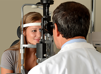
The layers of the cornea, iris, lens, and retina of the eye are examined in detail using a slit lamp, a device that is formed by a combination of many different devices. Thanks to this method, it is possible to determine the symptoms of almost all eye diseases.
-
Fundus examination
It is carried out with the use of dilating pupil drops. Thus, a more detailed examination of the lens and retina is possible. For this method, which examines the internal structures of the eye, it takes 20-30 minutes before the drops begin to act. In children, with the help of pupil enlargement, hidden visual defects can be detected.
With this examination, carried out using pupil dilation drops, symptoms of retinal detachment, glaucoma, hypertension, brain tumor, and various other diseases can be detected.
A thorough eye examination takes at least 30 minutes. During the examination, an examination of the fundus with the use of drops is necessary.
Prepared by the Dünyagöz Hospital Editorial Board.
*Page content is for informational purposes only. Consult your doctor for diagnosis and treatment
Last Update Date: 12.05.2023


